You are here
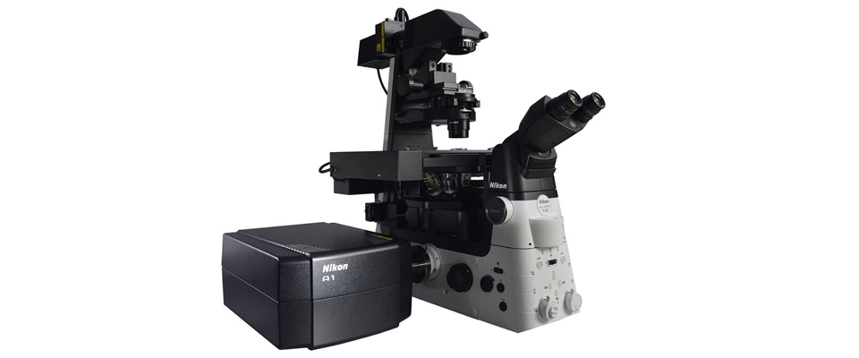
Nikon A1R HD25 Confocal Microscope
Advantages
Confocal microscopy enables acquisition of 2D or 3D fluorescence images as well as time-lapse images by collecting emission light only from the focus of a laser beam. Excitation light generates fluorescence throughout the specimen and out-of-focus light is eliminated by a pinhole. This microscopy can be used for imaging multi labeled structures with submicron resolution in live or fixed tissue. The best quality is achieved close to the surface of the sample (<200 μm). However, with the advancement of tissue processing, large cleared pieces could be imaged (up the entire mouse brain or spinal cord).
Description
Nikon A1R upright confocal microscope is equipped with Tokai Hit environmental chamber to control temperature and gas. This lightweight chamber is designed to be installed on Nikon Piezo or xy motorized stages. Transmitted light DIC pictures are combined with fluorescent images in the same software. The setup combines four lasers (405 nm, 488 nm, 561 nm, and 640 nm) and four detectors (2 GaAsP in green/red fluorescence and 2 PMT for blue/UV and far red fluorescence) for up to five individually regulated channels. A1-DUS spectral detector unit in Nikon’s system allows simultaneous excitation with 4 lasers and can be used to separate 32 bands of fluorescent spectra, allowing real time unmixing of closely overlapping emission profiles of multiple fluorescent probes (cell cultures) as well as removal of autofluorescence (large spinal cord and brain samples). Switchable between resonant and nonresonant mode within the same program for optimal scanning speeds. With a high-speed imaging capability up to 720 fps and 25 mm FOV, the A1R HD25 can dramatically increase imaging throughput. Using resonant scanning a higher fluorescence quantum yield and reduced photobleaching are achieved. In addition, powerful “Denoise” processing module can further improve image quality. Nikon’s proprietary Perfect Focus System (PFS) corrects for focus drift caused by temperature changes or mechanical disturbances. The PFS allows for the maintenance of focus in longitudinal measurements. This microscope can do spectral detection, time-lase imaging in living cells, 3D imaging, high content imaging.
The microscope has the following objectives: 10x (air, WD 4, NA 0.45, FOV 25 mm), 10x ( glycerin, WD 5.5, NA 0.5, FOV 25 mm), 20x (air, WD 1, NA 0.75, FOV 25 mm), 20x (water, WD 0.95, NA 0.95, FOV 22 mm), 40x (water, WD 0.17-0.25, NA, 0.95, FOV 25 mm), 40x (oil, WD 0.2, NA, 1.3, FOV 25 mm), 60x (oil, WD 0.13 NA 1.4, FOV 25). The microscope will work with any Nikon objectives. Multiple digital zoom options are available.
Generally, data analysis is conducted on a different computer using Fiji or Imaris.
After individual consultation and training sessions, researchers are given access to schedule instrument time through the institutional online booking system “booked”. Assistance with imaging and experimental design is available during normal business hours.