You are here
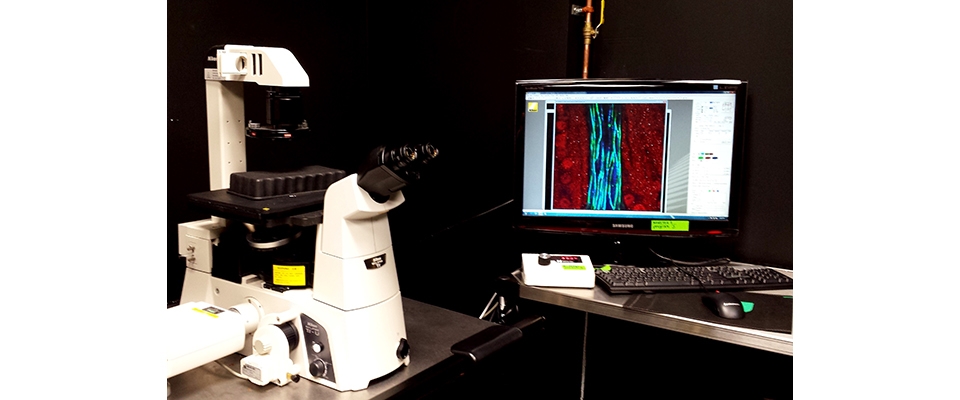
C1 Nikon Confocal Microscope
Advantages
Confocal microscopy enables acquisition of 2D or 3D fluorescence images as well as time-lapse images by collecting emission light only from the focus of a laser beam. Excitation light generates fluorescence throughout the specimen and out-of-focus light is eliminated by a pinhole. This microscopy can be used for imaging multi labeled structures with submicron resolution in live or fixed tissue. The best quality is achieved close to the surface of the sample (<200 μm).
Description
Our confocal microscope has three lasers (488 nm, 561 nm, and 633 nm) which can be used simultaneously or sequentially. The acquisition is relatively slow with ~1 fps rate at 1024x1024 pixels.
The microscope has the following objectives: 4x (air, WD 15.7, NA 0.2), 10x ( air, WD 10.5, NA 0.25), 10x (air, WD 4, NA 0.45), 20x (water, WD 0.35, NA 0.75), 40x (water, WD 2, NA, 0.8), 60x (water, WD 0.22 NA 1.2), 60x (oil, WD 0.13, NA 1.4), and 100x (oil, WD 0.13, NA 1.4). Additional 1.5 optical and multiple digital zoom options are available.
There is a port to attach a camera for imaging in epifluorescent or transmitted light. Camera-captured images will not be integrated into the confocal software. There is no tiling option (for automatic imaging of large cleared tissue samples) or an environmental chamber (for time-lapse imaging of living cell cultures).
Data analysis is typically conducted on a different computer using Fiji.
After individual consultation and training sessions, researchers are given access to schedule instrument time through the institutional online booking system “booked”. Assistance with imaging and experimental design is available during normal business hours.