You are here
Illuminating The Choroid Plexus-Cerebrospinal Fluid System
Speakers
Abstract
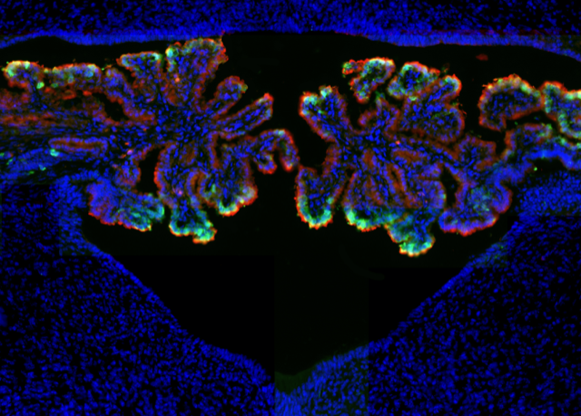
The choroid plexus (ChP) is a vital tissue located in each ventricle in the brain. The ChP is composed of two parallel sheets of epithelial cells with an intervening network of primarily non-neural cell types and vasculature. The ChP (1) produces cerebrospinal fluid (CSF) containing growth-promoting factors for the brain, (2) forms a blood-CSF barrier that gates communication between the central nervous system (CNS) and the systemic milieu, (3) regulates immune cell entry into the brain, and (4) offers an enticing framework for enhanced drug delivery. While ideally positioned to regulate brain function broadly from development to adulthood, the ChP network is surprisingly poorly understood compared to other neural and non-neural brain systems. Progress in understanding its role has been hindered in large part due to the lack of available tools for selectively accessing and controlling the ChP in vivo. We are developing a toolkit to enable a modern, system-level approach to ChP study in vivo. We recently used single cell sequencing to comprehensively classify and characterize the resident cells of the ChP. This new map enables the engineering of mouse driver lines for specific ChP cell types, allowing precise monitoring and control of each type. In parallel, we have been developing approaches to observe and control calcium activity patterns, detailed methods for 3D two-photon imaging of ChP cells, and methods for tracking the same cells across hours, days and weeks. We are using these tools to address questions regarding ChP functions in development, homeostasis, and in response to disease challenge.


