You are here
Uncovering Mechanisms of Neurodegeneration with Intravital Optical Imaging
Speakers
Abstract
The dynamics and logic of neuro-glio-vascular interactions in health and disease
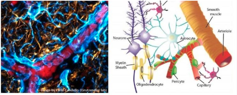
Our knowledge about the complex interplay between the various brain cells types (neuronal and non-neuronal) is still rudimentary. These interactions are disrupted in every neurological disorder. Our laboratory is interested in elucidating these multicellular and complex interactions that occur during brain pathogenesis. Recent innovations in live imaging and optical probes are allowing sophisticated interrogation of the structural and functional cellular changes that occur in pathological processes. Our goal is to develop and implement such methodologies for advancing our understanding of the physiology of different brain cells and how they interact in their native unperturbed microenvironment and during homeostatic perturbations. Our lab uses a variety of techniques including two-photon microscopy to repeatedly image individual neurons, glial cells (microglia, astrocytes, pericytes) and blood vessels over periods of up to months. We also developed a methodology for in vivo label free microscopy of myelin to study axonal myelination. This imaging-centric approach is combined with the use of viral vectors and in utero electroporation, optical sensors of cellular physiology, optogenetics, chemogenetics and genome editing techniques. Specific disease conditions that we are interested in include: Alzheimer’s, microvascular and myelin pathologies.


