You are here
Cellular and Molecular Characterization of Organ-Wide Neural Complexity and Dynamics
Speakers
Our nervous system is composed of many billions of neurons that form enormous amount of connections between each other and to the peripheral organs to regulate nearly all aspects of our physiological functions. Understanding the structural organization of the complex neural network is the key to unraveling how the nervous system regulates important body functions and controls complex animal behaviors. It is also the key to informing our understanding of the pathogenesis underlying neurological disorders. My research has been focused on understanding the conserved mechanism underlying the establishment of the vastly diversified neural connections. I have established robust in vivo and in vitro systems to address the central question: how do extending neurites precisely regulate their responses to complex environment during development to ensure accurate long-range projection. This will help us to understand how the neural circuitries may get disrupted in various neurodevelopmental disorders. It will also provide insights for how neural projections are dynamically modified under normal and diseased conditions later in the adult. To facilitate the analysis of the complex neuronal projections, which are often thin but extensive over large three-dimensional space, I have also developed advanced whole tissue clearing and imaging techniques (iDISCO, iDISCO+, Adipo-Clear, and the current “U.Clear” method) to visualize the entire neural morphologies in large intact organs. My platform is broadly applicable to enable systematic profiling of cellular organizations and functions in a more global scale. This will promote unbiased discoveries in brain research and beyond.

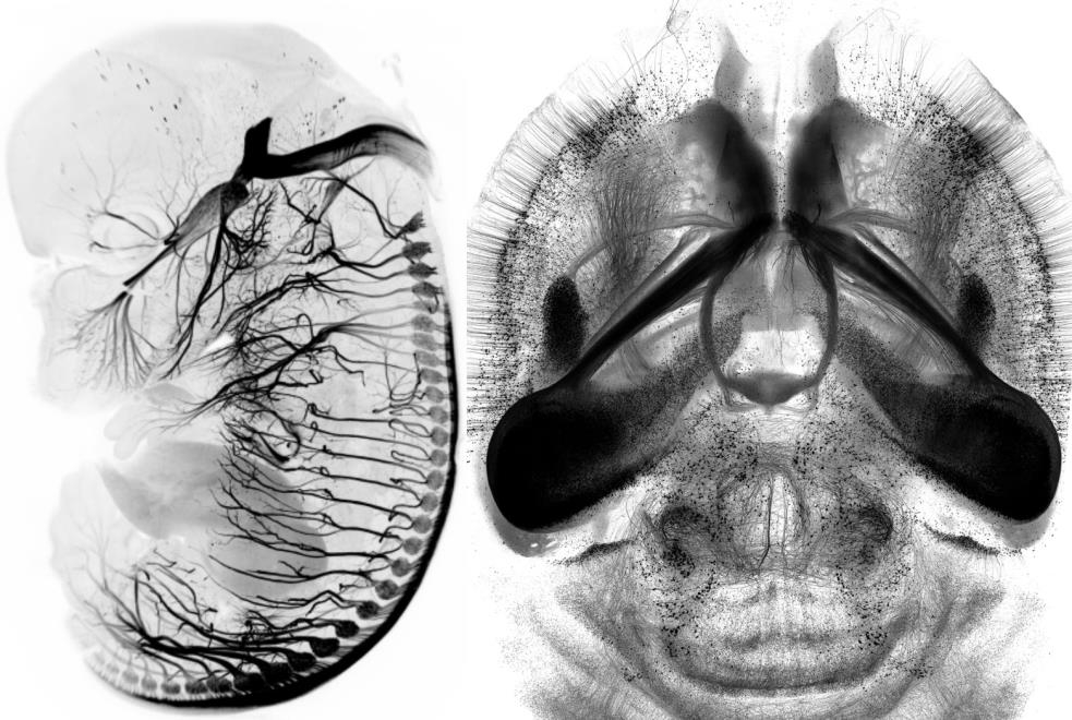
For the diagram, one represents the two areas that I have been working on (neurodevelopment and brain circuitry imaging): Left, a mouse embryo labeling the TrkA+ sensory pathway; Right, an adult mouse brain with Thy1-GFP reporter.

