You are here
Transcriptomic Responses of the Retina and Optic Nerve Head to Intraocular Pressure Elevation in the Human Eye
Speakers

Abstract
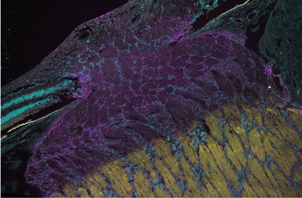
Glaucoma is a leading cause of blindness worldwide driven by progressive degeneration of retinal ganglion cells (RGCs). While age and elevated intraocular pressure (IOP) are key risk factors, lowering IOP remains the only established treatment. Although animal models have been instrumental in studying ocular perfusion, retinal function, and cellular responses to IOP elevation, they differ significantly from humans in immune responses and optic nerve head (ONH) architecture. This work leverages the Living Eye Project, which provides experimental access to human eyes in vivo in research-consented brain-dead organ donors prior to organ procurement, followed by ex vivo molecular analysis of the same eyes. In vivo assessments include retinal function using electroretinography (ERG), structural imaging with optical coherence tomography (OCT), and blood flow analysis using OCT angiography (OCT/A). Ex vivo, we examine ONH changes following IOP elevation including axoplasmic stasis and spatial transcriptomic responses. We hypothesize that elevation of IOP causes deformation of the ONH, which drives mechanosensitive mechanisms within the lamina cribrosa (LC) and peripapillary sclera to initiate remodeling within the LC. We are also able to use RNA-in situ hybridization and spatial transcriptomics at the macula to evaluate early transcriptomic responses of RGCs and their milieu to IOP elevation. We hypothesize that acute, subischemic elevations in IOP are capable of inducing an inflammatory and senescent phenotype in the inner retina, sensitizing RGCs to further injury. This work aims to identify early molecular events that render RGCs vulnerable, ultimately guiding the development of neuroprotective therapies for glauco