You are here
Structural and Functional Imaging Core
The Structural and Functional Imaging Core at the Burke Neurological Institute is a shared resource facility that provides state-of-the-art instruments, services, consultation, and assistance to all users at the Institute. Its instruments include widefield, laser scanning confocal and multi-photon, and laser capture microscopes along with an image processing workstation with software for 2D and 3D image analysis and quantification. The Imaging Core offers experimental services and consultation for sample preparation, training on the microscopes, providing imaging support, and assistance with analysis strategies. The Core also offers consultation and educational resources for designing new imaging experiments, and for testing hypotheses for publications or funding opportunity.
In 2020, with funding support of more than a half a million dollars from the National Institutes of Health Research Infrastructure Programs, the Imaging Core acquired Nikon’s latest high-end confocal microscope. With its large working field of view and high-speed, high-resolution resonant scanner, the Nikon microscope provided much-needed infrastructure for large tissue 3D imaging and key support to advance several NIH projects at our Institute. Moreover, Nikon microscope’s industry leading technologies for live cell imaging has allowed users to develop new experiments and expand the limits of our scientific research. Equipping our Imaging Core facility with state-of-the-art technology creates a central hub for the Institute where hypotheses risen by our research labs are propelled into experiments that pave the way to tomorrow’s discoveries.
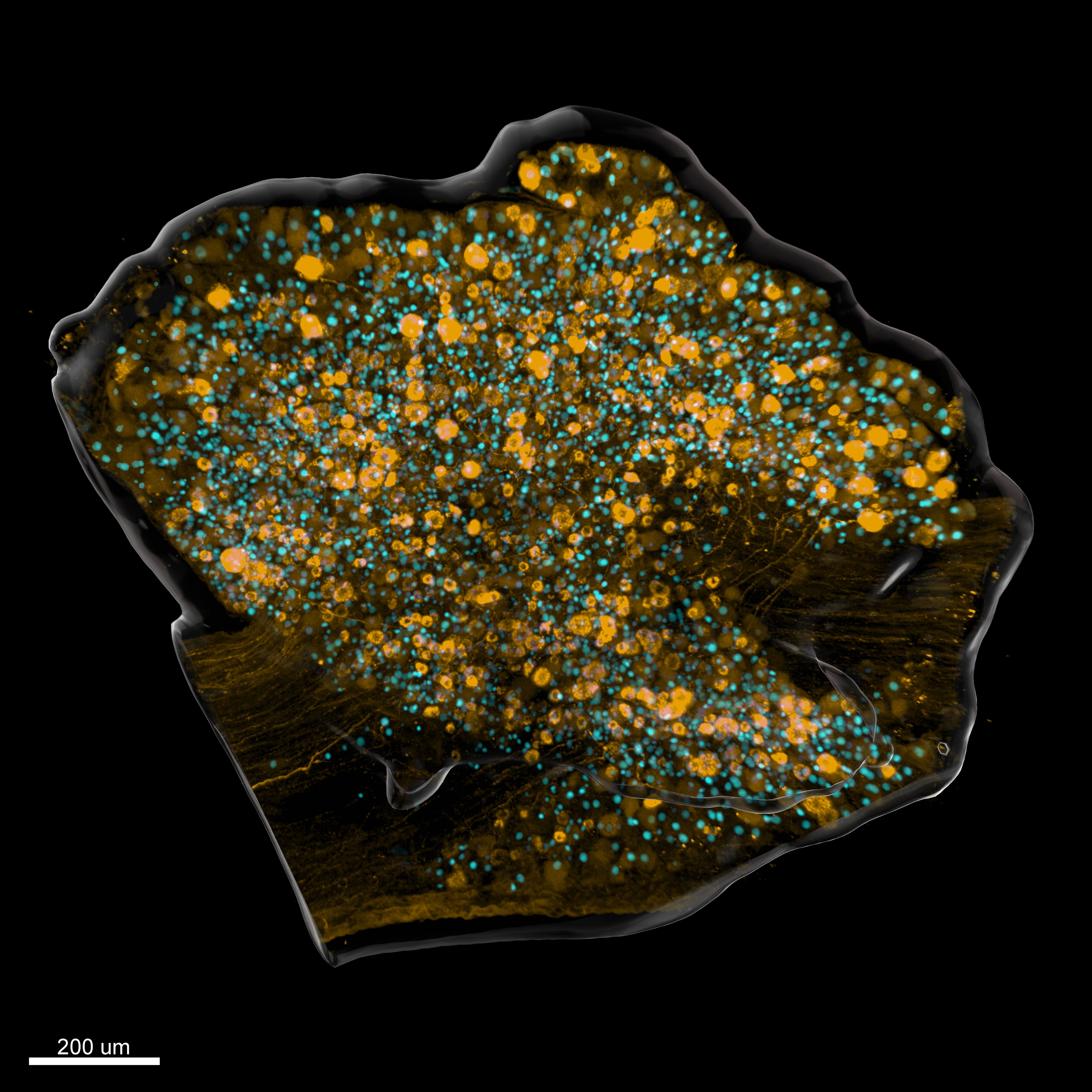
Figure 1. Regeneration associated gene activation in a whole-lumbar dorsal root ganglion. Three-dimensional surface rendering of mouse dorsal root ganglion seven days after sciatic nerve injury, showing the activation of the transcription factor ATF3 (cyan) and the calcitonin generelated peptide (CGRP) positive neurons (yellow). Courtesy of J. Sebastián Jara, Ph.D. of the Hollis Lab.
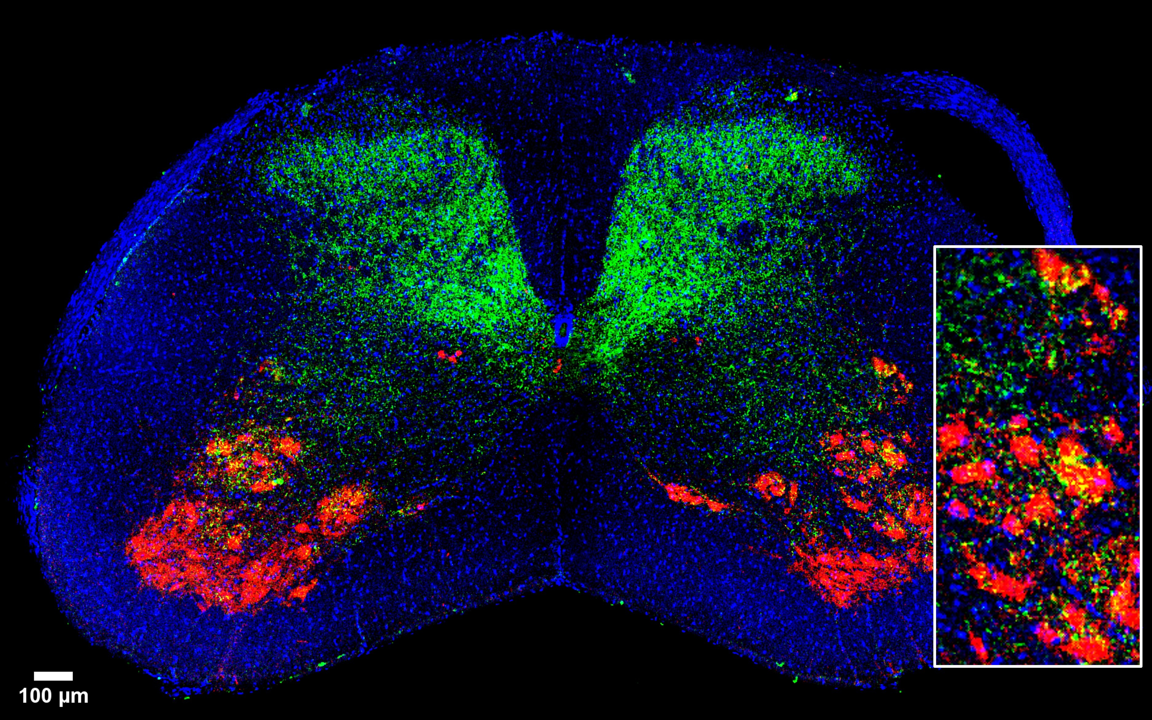
Figure 2. Spinal cord section C6 from a PV-cre mouse stained with vGlut1 (green), chAT (red), and DAPI (blue). Colocalization of vGlut1 and chAT indicates presence of proprioceptive sensory neuron synapses in motor neurons. Courtesy of Agustin Letelier of the Yoshida Lab.